SONOSITE X-PORTE
SonoSite X-Porte Features
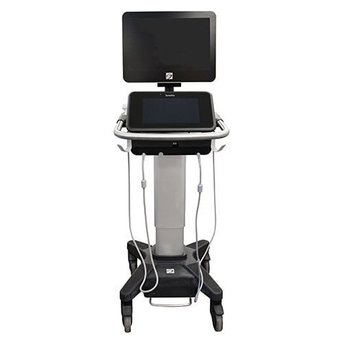
The SonoSite X-Porte is a point-of-care ultrasound system equipped with Extreme Definition Imaging (XDI) technology. This shared server ultrasound system is versatile, providing imaging for a wide range of procedures, including vascular, venous, cardiac, small parts, OB/GYN, nerve, superficial, MSK, and abdominal applications. Designed to be compact and portable, the X-Porte easily maneuvers around exam tables and offers adjustability for convenience. It features a 12-inch touchscreen control panel and a 19-inch HD monitor, delivering high-definition images. The monitor can also be tilted and rotated for optimal viewing angles.
- Touch Screen Interface
- Internal library of 87 tutorials
- 12-inch Touch screen Control panel
- Internal DVR that can record 60 minutes of scanning
- Compatible with at least 14 SonoSite transducers
- Extreme Definition Imaging (XDI) beam-forming technology
SonoSite X-Porte Specifications
Dimensions
- Height (max):64 in (162.6 cm)
- Height (min):42.2 in (107.2 cm)
- Height Adjustment: 9 in (22.9 cm)
- Width: 21.2 in (53.8 cm)
- Length: 26.4 in (67.1 cm)
Touch Panel
- Capacitive display Type
- Diagonal: 12.1 in (30.7)
- Tilt Adjustment: 7.3 in (18.5 cm)
- Side to Side Turning: +/- 9 degrees from center
Display
- Diagonal: 19in (48.3 cm)
- Screen Size: 1280 x 800
- Image Size: 800 x 600
- Monitor Tilt: 5 degrees tilting forward from vertical, 20 degrees tilting back from vertical
Electrical
- Power input (stand version): 100-240V~6.0A Max, 50-60Hz
- Power output (stand Version): 24 VDC, 11.5 A Max (output not exceeding 275 watts)
- Power Input (Desktop Version): 100-240 V~3.4A- 1.4A, 50-60Hz
- Power Output (Desktop Version): 24 VDC, 6.25A Max (Combined Output not exceeding 10 watts)
- Batteries (Stand Version): 3 Lithium-ion batteries (385Wh total)
- Battery Use Time: 1.0 hours, 3 days on idle
- Battery Charge Time: 2.5 hours
- Battery Life: 3-6 years
Imaging Modes
- 2D, Broadband Imaging
- Tissue Harmonic Imaging (P21xp, C60xp)
- Pulse Inversion Harmonic Imaging
- M-mode
- Velocity Color Doppler
- Color Power Doppler
- Pulsed Wave Doppler
- Pulsed Wave Tissue Doppler
- Continuous Wave Doppler, ECG
Image Processing
- Extreme Definition Imaging (XDI)
- SonoAdapt Tissue Optimization
- SonoHD2 Imaging Technology
- Dual Imaging
- Dual Color Imaging
- SonoMB Multibeam Technology
- AutoGain
- AutoGain Brightness Adjust
- Restore Default Gains
- Dynamic Range
- Duplex Imaging
- 8x Zoom Capability
- Post Processing: Dynamic Range, Zoom
- 2D Image Optimization: Average and Difficult
- Overall Gain, Near and Far Field Gain Control
- Color and Doppler Flow Optimization (low, medium, high)
- Color Variance Mode
- 2D Reduced Imaging Sector
User Interface and Programmable Controls
- Capacitive Touch Screen
- Multi-touch gestures for system controls Configurable User Interface: Start Screen, More Controls, Programmable Keys, System Parameters Clinical Display Information
- Programmable Keys (9): Functions: Show/Hide, End Exam, Reset Gain to
Default Values, Print, Save Image, Save Video Clip, AutoGain, Calcs, None
- Configurable Start Screen: Start, Scanning, Transducer/Exam Selection, Patient Information
- Virtual QWERTY Keyboard for annotation
- User defined exam types (up to five exam types for each exam type/transducer combination). For example, you can define five different exam types for Abdomen on P21xp transducer and five exam types for Abdomen on the C60xp transducer.
- Image Acquisition Keys: Save, Review, Report, Video Clip Store, Video Clip Edit, DVR
- Labeling of saved images
- Display formats for Duplex Imaging: 1/3 and 2/3, ½ and ½, 2/3 and 1/3, side by side and full-screen duplex
- Doppler Controls: angle, steer, scale, baseline, sample volume, gain and volume
Measurements
- 2D: Distance – 8 measurements, Ellipse, Manual Trace Volume, Target Depth, Bladder Volume
- Doppler: Velocity measurements, Pressure Gradient, Elapsed Time, Acceleration, Heart Rate, Resistive Index, Systolic/Diastolic Ratio, Measurements can be traced manually or automatically.
- Automatic trace results (determined by exam type): Velocity Time Integral, Peak Velocity, Mean Pressure Gradient, Mean Velocity on Peak Trace, Press Gradient, Cardiac Output, Peak Systolic Velocity, Time Average Mean, Systolic/Diastolic Ratio, Pulsatility Index, End Diastolic Velocity, Acceleration Time, Resistive Index, Time Average Peak, Gate Depth, Heart Rate.
- M-mode: All points guided workflow, distance and time measurements, Heart Rate
- Editable results data sheets and reports
Sonosite X-Porte
- Product Code: SS678853
SONOSITE X-PORTE
SonoSite X-Porte Features
The SonoSite X-Porte is a point-of-care ultrasound system equipped with Extreme Definition Imaging (XDI) technology. This shared server ultrasound system is versatile, providing imaging for a wide range of procedures, including vascular, venous, cardiac, small parts, OB/GYN, nerve, superficial, MSK, and abdominal applications. Designed to be compact and portable, the X-Porte easily maneuvers around exam tables and offers adjustability for convenience. It features a 12-inch touchscreen control panel and a 19-inch HD monitor, delivering high-definition images. The monitor can also be tilted and rotated for optimal viewing angles.
- Touch Screen Interface
- Internal library of 87 tutorials
- 12-inch Touch screen Control panel
- Internal DVR that can record 60 minutes of scanning
- Compatible with at least 14 SonoSite transducers
- Extreme Definition Imaging (XDI) beam-forming technology
SonoSite X-Porte Specifications
Dimensions
- Height (max):64 in (162.6 cm)
- Height (min):42.2 in (107.2 cm)
- Height Adjustment: 9 in (22.9 cm)
- Width: 21.2 in (53.8 cm)
- Length: 26.4 in (67.1 cm)
Touch Panel
- Capacitive display Type
- Diagonal: 12.1 in (30.7)
- Tilt Adjustment: 7.3 in (18.5 cm)
- Side to Side Turning: +/- 9 degrees from center
Display
- Diagonal: 19in (48.3 cm)
- Screen Size: 1280 x 800
- Image Size: 800 x 600
- Monitor Tilt: 5 degrees tilting forward from vertical, 20 degrees tilting back from vertical
Electrical
- Power input (stand version): 100-240V~6.0A Max, 50-60Hz
- Power output (stand Version): 24 VDC, 11.5 A Max (output not exceeding 275 watts)
- Power Input (Desktop Version): 100-240 V~3.4A- 1.4A, 50-60Hz
- Power Output (Desktop Version): 24 VDC, 6.25A Max (Combined Output not exceeding 10 watts)
- Batteries (Stand Version): 3 Lithium-ion batteries (385Wh total)
- Battery Use Time: 1.0 hours, 3 days on idle
- Battery Charge Time: 2.5 hours
- Battery Life: 3-6 years
Imaging Modes
- 2D, Broadband Imaging
- Tissue Harmonic Imaging (P21xp, C60xp)
- Pulse Inversion Harmonic Imaging
- M-mode
- Velocity Color Doppler
- Color Power Doppler
- Pulsed Wave Doppler
- Pulsed Wave Tissue Doppler
- Continuous Wave Doppler, ECG
Image Processing
- Extreme Definition Imaging (XDI)
- SonoAdapt Tissue Optimization
- SonoHD2 Imaging Technology
- Dual Imaging
- Dual Color Imaging
- SonoMB Multibeam Technology
- AutoGain
- AutoGain Brightness Adjust
- Restore Default Gains
- Dynamic Range
- Duplex Imaging
- 8x Zoom Capability
- Post Processing: Dynamic Range, Zoom
- 2D Image Optimization: Average and Difficult
- Overall Gain, Near and Far Field Gain Control
- Color and Doppler Flow Optimization (low, medium, high)
- Color Variance Mode
- 2D Reduced Imaging Sector
User Interface and Programmable Controls
- Capacitive Touch Screen
- Multi-touch gestures for system controls Configurable User Interface: Start Screen, More Controls, Programmable Keys, System Parameters Clinical Display Information
- Programmable Keys (9): Functions: Show/Hide, End Exam, Reset Gain to
Default Values, Print, Save Image, Save Video Clip, AutoGain, Calcs, None
- Configurable Start Screen: Start, Scanning, Transducer/Exam Selection, Patient Information
- Virtual QWERTY Keyboard for annotation
- User defined exam types (up to five exam types for each exam type/transducer combination). For example, you can define five different exam types for Abdomen on P21xp transducer and five exam types for Abdomen on the C60xp transducer.
- Image Acquisition Keys: Save, Review, Report, Video Clip Store, Video Clip Edit, DVR
- Labeling of saved images
- Display formats for Duplex Imaging: 1/3 and 2/3, ½ and ½, 2/3 and 1/3, side by side and full-screen duplex
- Doppler Controls: angle, steer, scale, baseline, sample volume, gain and volume
Measurements
- 2D: Distance – 8 measurements, Ellipse, Manual Trace Volume, Target Depth, Bladder Volume
- Doppler: Velocity measurements, Pressure Gradient, Elapsed Time, Acceleration, Heart Rate, Resistive Index, Systolic/Diastolic Ratio, Measurements can be traced manually or automatically.
- Automatic trace results (determined by exam type): Velocity Time Integral, Peak Velocity, Mean Pressure Gradient, Mean Velocity on Peak Trace, Press Gradient, Cardiac Output, Peak Systolic Velocity, Time Average Mean, Systolic/Diastolic Ratio, Pulsatility Index, End Diastolic Velocity, Acceleration Time, Resistive Index, Time Average Peak, Gate Depth, Heart Rate.
- M-mode: All points guided workflow, distance and time measurements, Heart Rate
- Editable results data sheets and reports
- Brand: Sonosite
- Availability: In Stock


